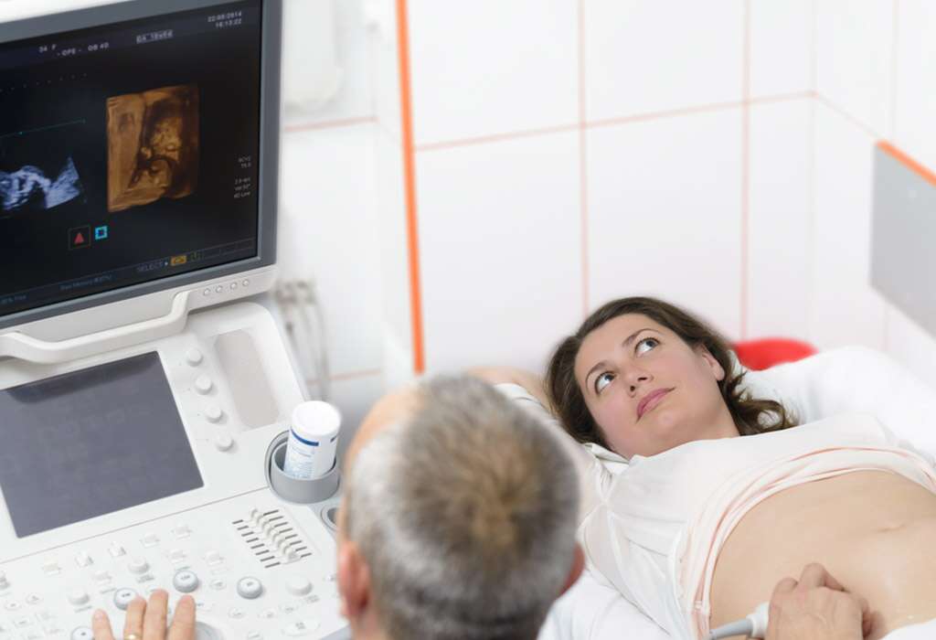
Most pregnant women are eager to get their ultrasounds and can’t wait for it to happen; the purpose of these belly scans is to provide expecting mothers with the first glimpse of their unborn baby. It is a very emotional time for the woman and her family as well because they get to see the baby in the womb for the very first time. Since the late 1950s, obstetricians have been using ultrasounds to provide a confirmation of pregnancy, establish the size of the fetus, the gestational age, and figure out the sex and also to keep track of the placement and development of the baby. They are also useful for highlighting any problems concerning the fetus.
The schedule of the ultrasounds is typically provided by the obstetricians. Typically, the first ultrasound is performed after seven weeks for confirmation of the pregnancy. The gender scan and anatomy of the baby is evaluated through an ultrasound that’s performed between 160 to 20 weeks and it also checks the baby for any abnormalities. When 32 weeks have passed in the pregnancy, the baby scan is performed to judge the growth and feet size of the baby. If there is any danger of fetal abnormalities or mother goes beyond the due date or is at risk, additional pregnancy scan can also be performed.
An ultrasound scan may also be performed by some doctors between 11 to 14 weeks for checking for symptoms of Down syndrome. There are different types of ultrasounds that can be performed during pregnancy. In early pregnancy, transvaginal scans may be performed in which sonogram images are created by inserting probe transducers vaginally. 2-Dimensional images of the fetus are provided by standard or traditional scans in which a transducer is glided over the belly of the pregnant woman.
With technological advancement, there has been an introduction of advanced methods of performing pregnancy scans, which use high-end equipment. For instance, blood flow and blood pressure are measured in Doppler ultrasounds by bouncing off the waves of the ultrasound off the blood cells. Likewise, nowadays, there are 3D and 4D scans also being performed, but they also need proper equipment, which people can find at Baby Moments. Here expectant mothers and other family members can go together to undergo an ultrasound scan to catch a glimpse of their newest family member.
At Baby Moments, a 3D ultrasound is performed in the same way as 2D ultrasound, with the use of sound waves. The difference is that it doesn’t produce a 2D lateral reading; instead, transducers are used in 3D scans for taking pictures of the fetus from several angles. These pictures are then merged into a computer to form 3D images and provide a better picture than 2D scans. Also known as dynamic 3D scans, 4D scans are similar to a DVD as they allow parents to see the facial expressions of the baby when it moves around in the womb. They are performed between 28 to 32 weeks of the pregnancy and people can visit Baby Moments to make this time memorable for them. We cover Berkshire Wiltshire and Oxfordshire.